Last updated on April 15 2024
Description
Proximal Femoral Osteotomy (PFO) is a joint-sparing procedure that relies on maintaining the biologic integrity of the femoral head. This type of osteotomy is used in the treatment of hip fracture nonunions and malunions and in cases of congenital and acquired hip deformities. Deformities typically include a varus or valgus neck-shaft angle (CCD), rotational malalignments, and leg-length discrepancy in any combination. Consequently, the goal of PFO is to correct the deformity, by realigning the hip and lower extremities.
During this osteotomy, a wedge of bone is removed to realign and reposition the femoral head.
- Coxa Vara: A decreased neck-shaft angle is called coxa vara or varus alignment, CCD < 120°.
- Normal CCD (adult) ranges from 120º to 135º (most studies use a mean value of 130°).
- Coxa Valga: An increased neck-shaft angle is called coxa valga or valgus alignment, CCD > 135°.
Measurements
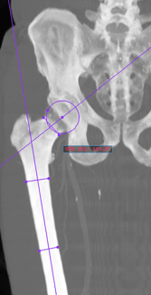
The measurements made by the procedure are displayed in the image:
- Centrum-Collum-Diaphyseal (CCD): the angle between the Femoral Shaft Axis (FSA) and the Femoral Neck Axis (FNA).
Auxiliary references
To show this procedure, PeekMed® needs additional references:
- Femoral Shaft Axis (FSA): the line that passes through the center of the femoral shaft;
- Femoral Neck Axis (FNA): the line that passes through the center of the femoral head and neck.
How to perform
After selecting this procedure in the sidebar, you must mark the points to conclude it. To see the caption of each point you need to click on this button .
Beware: you can change the position of the points at this stage or later. To do this, simply click on each handle with the left mouse button and move them to the most suitable position.
For advanced configuration, please follow the instructions below.
Advanced Configuration
After the correct positioning of the PFO procedure, if a deformity is identified, it is possible to correct it automatically using this option on Object edition, to restore the patient's anatomical alignment. Based on the value obtained (CCD), the procedure configuration will show the type of deformity correction that can be performed (Varus or Valgus).
After clicking “Next”, it is possible to cut a fragment or to select an existing fragment of the femur, taking into consideration if a Conventional image, CR, CT (segmented or not-segmented), or MRI is being used.

|

|

|
After determining if the patient has a deformity, the femur fragment should be placed correctly in the acetabulum and a second set of points should be marked, to define the placement of the resection line:
Point 9 - Mark a point at the medial edge of the $(side) femur to define the resection line.
Point 10 - Mark a point at the lateral edge of the $(side) femur to define the resection line.
After marking the points, it is possible to perform the Osteotomy by clicking on Finish.

|

|
|
